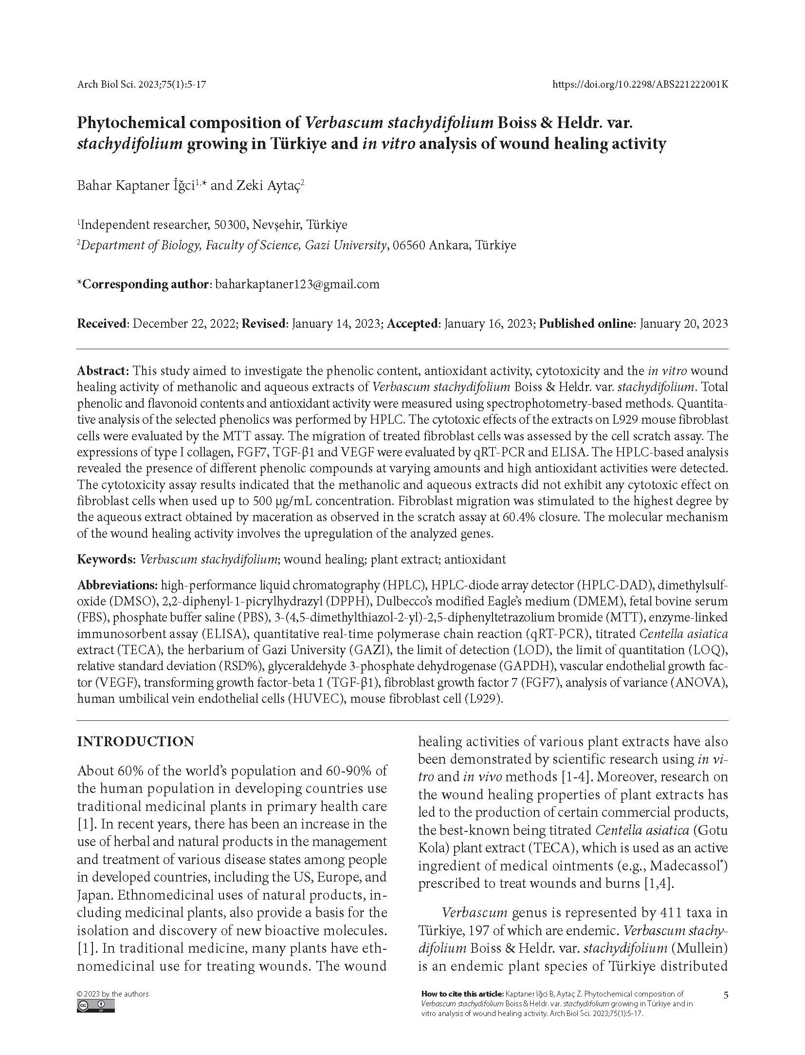
Phytochemical composition of Verbascum stachydifolium Boiss & Heldr. var. stachydifolium growing in Türkiye and in vitro analysis of wound healing activity
DOI:
https://doi.org/10.2298/ABS221222001KKeywords:
Verbascum stachydifolium, wound healing, plant extract, antioxidantAbstract
Paper description:
- The aim of this study is to support alternative treatment methods that will provide wound healing and to investigate the molecular and phytochemical characteristics of Verbascum stachydifolium
- The scratch assay was used to assess wound healing activity in vitro, qRT-PCR and ELISA were performed to evaluate the expression of selected biomarkers, HPLC was used to determine the phytochemical components of stachydifolium.
- stachydifolium exhibited a wound healing potential by increasing the expression of collagen and cytokines FGF7, TGF-b1 and VEGF. The plant also possesses antioxidant activity.
- stachydifolium emerges as a potential source for plant-based wound healing preparations.
Abstract: This study aimed to investigate the phenolic content, antioxidant activity, cytotoxicity and the in vitro wound healing activity of methanolic and aqueous extracts of Verbascum stachydifolium Boiss & Heldr. var. stachydifolium. Total phenolic and flavonoid contents and antioxidant activity were measured using spectrophotometry-based methods. Quantitative analysis of the selected phenolics was performed by HPLC. The cytotoxic effects of the extracts on L929 mouse fibroblast cells were evaluated by the MTT assay. The migration of treated fibroblast cells was assessed by the cell scratch assay. The expressions of type I collagen, FGF7, TGF-β1 and VEGF were evaluated by qRT-PCR and ELISA. The HPLC-based analysis revealed the presence of different phenolic compounds at varying amounts and high antioxidant activities were detected. The cytotoxicity assay results indicated that the methanolic and aqueous extracts did not exhibit any cytotoxic effect on fibroblast cells when used up to 500 µg/mL concentration. Fibroblast migration was stimulated to the highest degree by the aqueous extract obtained by maceration as observed in the scratch assay at 60.4% closure. The molecular mechanism of the wound healing activity involves the upregulation of the analyzed genes.
Downloads
References
Agyare C, Bekoe EO, Boakye YD, Dapaah SO, Appiah T, Bekoe SO. Medicinal Plants and Natural Products with Demonstrated Wound Healing Properties. In: Alexandrescu V, editor. Wound Healing - New insights into Ancient Challenges. London: InTechOpen; 2016. p. 487-535. https://doi.org/10.5772/63574
Süntar I, Akkol EK, Keleş H, Oktem A, Başer KHC, Yeşilada E. A novel wound healing ointment: A formulation of Hypericum perforatum oil and sage and oregano essential oils based on traditional Turkish knowledge. J Ethnopharmacol. 2011;134:89-96. https://doi.org/10.1016/j.jep.2010.11.061
Süntar I, Tatlı II, Küpeli Akkol E, Keleş H, Kahraman Ç, Akdemir Z. An ethnopharmacological study on Verbascum species: From conventional wound healing use to scientific verification. J Ethnopharmacol. 2010;132:408-13. https://doi.org/10.1016/j.jep.2010.08.004
Maver T, Maver U, Stana Kleinschek K, Smrke DM, Kreft S. A review of herbal medicines in wound healing. Int J Dermatol. 2015;54:740-51. https://doi.org/10.1111/ijd.12766
Güner Adil, Aslan S, Ekim T, Babaç T. List of Turkish Flora (Vascular Plants). Nezahat Gökyiğit Botanical Garden and Flora Research Society; 2012.
Ozkan G, Kamiloglu S, Ozdal T, Boyacioglu D, Capanoglu E. Potential use of Turkish medicinal plants in the treatment of various diseases. Molecules. 2016;21:1-32. https://doi.org/10.3390/molecules21030257
Demirci S, Doğan A, Demirci Y, Şahin F. In vitro wound healing activity of methanol extract of Verbascum speciosum. Int J Appl Res Nat Prod. 2014;7:37-44.
Akdemir Z, Kahraman Ç, Tatlı II, Küpeli Akkol E, Süntar I, Keles H. Bioassay-guided isolation of anti-inflammatory, antinociceptive and wound healer glycosides from the flowers of Verbascum mucronatum Lam. J Ethnopharmacol. 2011;136:436-43. https://doi.org/10.1016/j.jep.2010.05.059
Singleton VL, Orthofer R, Lamuela-Raventós RM. Analysis of total phenols and other oxidation substrates and antioxidants by means of folin-ciocalteu reagent. Methods Enzymol. 1999;299:152-78. https://doi.org/10.1016/S0076-6879(99)99017-1
Kaptaner İğci B, Aytaç Z. An investigation on the in vitro wound healing activity and phytochemical composition of Hypericum pseudolaeve Robson growing in Turkey. Turk J Pharm Sci. 2020;17:610-9. https://doi.org/10.4274/tjps.galenos.2019.80037
Aliyu AB, Ibrahim MA, Musa AM, Musa AO, Kiplimo JJ, Oyewale AO. Free radical scavenging and total antioxidant capacity of root extracts of Anchomanes difformis ENGL. (ARACEAE).Acta Pol Pharm. 2013;70:115-21.
Blois MS. Antioxidant determinations by the use of a stable free radical. Nature. 1958;181:1199-200. https://doi.org/10.1038/1811199a0
Magnusson B, Örnemark U. The Fitness for Purpose of Analytical Methods A Laboratory Guide to Method Validation and Related Topics. 2nd ed. Teddington: Eurachem; 2014. 70 p.
Mosmann T. Rapid colorimetric assay for cellular growth and survival: application to proliferation and cytotoxicity assays. J Immunol Methods. 1983;65:55-63. https://doi.org/10.1016/0022-1759(83)90303-4
Felice F, Zambito Y, Belardinelli E, Fabiano A, Santoni T, di Stefano R. Effect of different chitosan derivatives on in vitro scratch wound assay: a comparative study. Int J Biol Macromol. 2015;76:236-41. https://doi.org/10.1016/j.ijbiomac.2015.02.041
Livak KJ, Schmittgen TD. Analysis of relative gene expression data using real-time quantitative PCR and the 2(-Delta Delta C(T)) Method. Methods. 2001;25:402-8. https://doi.org/10.1006/meth.2001.1262
Agar OT, Dikmen M, Ozturk N, Yilmaz MA, Temel H, Turkmenoglu FP. Comparative studies on phenolic composition, antioxidant, wound healing and cytotoxic activities of selected Achillea L. species growing in Turkey. Molecules. 2015;20:17976-8000. https://doi.org/10.3390/molecules201017976
Alali FQ, Tawaha K, El-Elimat T, Syouf M, El-Fayad M, Abulaila K, Nielsen SJ, Wheaton WD, Falkinham JO. Antioxidant activity and total phenolic content of aqueous and methanolic extracts of Jordanian plants: an ICBG project. Nat Prod Res. 2007;21:1121-1131. https://doi.org/10.1080/14786410701590285
Mihailovic V, Kreft S, Benkovic ET, Ivanovic N. Chemical profile, antioxidant activity and stability in stimulated gastrointestinal tract model system of three Verbascum species. Ind Crops Prod. 2016;89:141-51. https://doi.org/10.1016/j.indcrop.2016.04.075
Akdemir ZŞ, Tatli II, Bedir E, Khan IA. Neolignan and phenylethanoid glycosides from Verbascum salviifolium boiss. Turk J Chem. 2004;28:621-8.
Kahraman C, Akdemir ZS, Tatli II. Promising Cytotoxic Activity Profile, Biological Activities and Phytochemical Screening of Verbascum L . Species. Med Aromat Plant Sci Biotechnol. 2012;6:63-75.
Turker AU, Camper ND, Gürel E. High-Performance Liquid Chromatographic Determination of a Saponin from Verbascum. Biotechnol Biotechnol Equip. 2004;18:54-9. https://doi.org/10.1080/13102818.2004.10819230
Akdemir ZS, Tatlı II, Bedir E, Khan IA. Antioxidant Flavonoids from Verbascum salviifolium Boiss. FABAD J Pharm Sci. 2003;28:71-5.
Grigore A, Colceru-Mihul S, Litescu S, Panteli M, Rasit I. Correlation between polyphenol content and anti-inflammatory activity of Verbascum phlomoides (mullein). Pharm Biol. 2013;51:925-9. https://doi.org/10.3109/13880209.2013.767361
Klimek B, Olszewska MA, Magdelena T. Simultaneous determination of flavonoids and phenylethanoids in the flowers of Verbascum densiflorum and V. phlomoides by high-performance liquid chromatography. Phytochem Anal. 2010;21:150-6. https://doi.org/10.1002/pca.1171
Ozcan B, Esen M, Caliskan M, Mothana RA, Cihan AC, Yolcu H. Antimicrobial and antioxidant activities of the various extracts of Verbascum pinetorum Boiss. O. Kuntze (Scrophulariaceae). Eur Rev Med Pharmacol Sci. 2011;15:900-5.
Caliskan B, Ozcan M, Yilmaz M. Antimicrobial and Antioxidant Activities of Various Extracts of Verbascum antiochium Boiss. (Scrophulariaceae). J Med Food. 2010;13:1147-52. https://doi.org/10.1089/jmf.2009.0213
Aligiannis N, Mitaku S, Tsitsa-Tsardis E, Catherine, Harvala Ioannis, Tsaknis Stavros L, Serko H. Methanolic Extract of Verbascum macrurum as a Source of Natural Preservatives against Oxidative Rancidity. J Agric Food Chem. 2003;51:7308-12. https://doi.org/10.1021/jf034528
Liu Z, Ren Z, Zhang J, Chuang C-C, Kandaswamy E, Zhou T, Zuo Li. Role of ROS and Nutritional Antioxidants in Human Diseases. Front Physiol. 2018;9:477. https://doi.org/10.3389/fphys.2018.00477
Skerget M, Kotnik P, Hadolin M, Ri A, Simoni M, Zeljko Kne. Phenols, proanthocyanidins , flavones and flavonols in some plant materials and their antioxidant activities. Food Chem. 2005;89:191-8. https://doi.org/10.1016/j.foodchem.2004.02.025
Tatlı İİ, Schuhly W, Akdemir ZS. Secondary Metabolites from Bioactive Methanolic Extract of Verbascum pycnostachyum Boiss. & Helder Flowers. Hacettepe Uni J Fac Pharm. 2007;27:23-32.
Armatu A, Bodırlau R, Nechıta CB, Nıculaua M, Teaca CA, Ichım M, Spiridon I. Characterization of biological active compounds from Verbascum phlomoides by chromatography techniques. I. Gas chromatography. Rom Biotechnol Lett. 2011;16:6297-304.
Tatlı İ, Akdemir Z. Cytotoxic Activity on Some Verbascum Species Growing in Turkey. Hacettepe Uni J Fac Pharm. 2006;26:77-85.
Fronza M, Heinzmann B, Hamburger M, Laufer S, Merfort I. Determination of the wound healing effect of Calendula extracts using the scratch assay with 3T3 fibroblasts. J Ethnopharmacol. 2009;126:463-7. https://doi.org/10.1016/j.jep.2009.09.014
Gill SE, Parks WC. Metalloproteinases and their inhibitors: Regulators of wound healing. Int J Biochem Cell Biol. 2008;40:1334-47. https://doi.org/10.1016/j.biocel.2007.10.024
Tsirogianni AK, Moutsopoulos NM, Moutsopoulos HM. Wound healing: Immunological aspects. Injury. 2006;37:S5-S12. https://doi.org/10.1016/j.injury.2006.02.035
Martin P. Wound Healing-Aiming for Perfect Skin Regeneration. Science. 1997;276:75-81. https://doi.org/10.1126/science.276.5309.75
Maquart F-X, Bellon G, Gillery P, Wegrowski Y, Borel J-P. Stimulation of Collagen Synthesis in Fibroblast Cultures by a Triterpene Extracted from Centella asiatica. Connect Tissue Res. 1990;24:107-20. https://doi.org/10.3109/03008209009152427
Barrientos S, Stojadinovic O, Golinko MS, Brem H, Tomic-Canic M. Growth factors and cytokines in wound healing. Wound Repair Regen. 2008;16:585-601. https://doi.org/10.1111/j.1524-475X.2008.00410.x
Niu J, Chang Z, Peng B, Xia Q, Lu W, Huang P, Tsao MS, Chiao PJ. Keratinocyte growth factor/fibroblast growth factor-7-regulated cell migration and invasion through activation of NF-kappaB transcription factors. J Biol Chem. 2007;282:6001-11. https://doi.org/10.1074/jbc.M606878200
Robson MC, Phillips TJ, Falanga V, Odenheimer DJ, Parish LC, Jensen JL, Steed DL. Randomized trial of topically applied repifermin (recombinant human keratinocyte growth factor-2) to accelerate wound healing in venous ulcers. Wound Repair Regen. 2001;9:347-52. https://doi.org/10.1046/j.1524-475x.2001.00347.x
Frank S, H bner G, Breier G, Longaker MT, Greenhalgh DG, Werner S. Regulation of Vascular Endothelial Growth Factor Expression in Cultured Keratinocytes. J Biol Chem. 1995;270:12607-13. https://doi.org/10.1074/jbc.270.21.12607
Ashcroft GS, Dodsworth J, Boxtel E Van, Tarnuzzer RW, Horan MA, Schultz GS, Ferguson MW. Estrogen accelerates cutaneous wound healing associated with an increase in TGF-β1 levels. Nat Med. 1997;3:1209-15. https://doi.org/10.1038/nm1197-1209
Lee H-S, Kooshesh F, Sauder D, Kondo S. Modulation of TGF-beta 1 production from human keratinocytes by UVB. Exp Dermatol. 1997;6:105-10. https://doi.org/10.1111/j.1600-0625.1997.tb00155.x
Wu L, Yu YL, Galiano RD, Roth SI, Mustoe TA. Macrophage Colony-Stimulating Factor Accelerates Wound Healing and Upregulates TGF-β1 mRNA Levels through Tissue Macrophages. J Surg Res 1997;72:162-9. https://doi.org/10.1006/jsre.1997.5178
Mani H, Sidhu GS, Kumari R, Gaddipati JP, Seth P, Maheshwari RK. Curcumin differentially regulates TGF-beta1, its receptors and nitric oxide synthase during impaired wound healing. Biofactors. 2002;16:29-43. https://doi.org/10.1002/biof.5520160104
Werner S, Krieg T, Smola H. Keratinocyte-Fibroblast Interactions in Wound Healing. Journal of Investigative Dermatology. 2007;127:998-1008. https://doi.org/10.1038/sj.jid.5700786
Chen L, Tredget EE, Wu PYG, Wu Y. Paracrine Factors of Mesenchymal Stem Cells Recruit Macrophages and Endothelial Lineage Cells and Enhance Wound Healing. PLoS One. 2008;3:1886. https://doi.org/10.1371/journal.pone.0001886
Anitua E, Pino A, Orive G. Plasma rich in growth factors promotes dermal fibroblast proliferation, migration and biosynthetic activity. J Wound Care. 2016;25:680-7. https://doi.org/10.12968/jowc.2016.25.11.680
Kane CJM, Hebda PA, Mansbridge JN, Hanawalt PC. Direct evidence for spatial and temporal regulation of transforming growth factor beta 1 expression during cutaneous wound healing. J Cell Physiol. 1991;148:157-73. https://doi.org/10.1002/jcp.1041480119
Colwell AS, Phan T-T, Kong W, Longaker MT, Lorenz PH. Hypertrophic scar fibroblasts have increased connective tissue growth factor expression after transforming growth factor-beta stimulation. Plast Reconstr Surg. 2005;116:1387-91. https://doi.org/10.1097/01.prs.0000182343.99694.28
Bao P, Kodra A, Tomic-Canic M, Golinko MS, Ehrlich HP, Brem H. The Role of Vascular Endothelial Growth Factor in Wound Healing. J Surg Res. 2009;153:347-58. https://doi.org/10.1016/j.jss.2008.04.023
Brown BLF, Yeo KT, Berse B, Yeo T, Senger DR, Dvorak HF. Expression of Vascular Permeability Factor (Vascular Endothelial Growth Factor) by Epidermal Keratinocytes during Wound Healing. J Exp Med. 1992;176:1376-9. https://doi.org/10.1084/jem.176.5.1375
Nissen NN, Polverini PJ, Koch AE, Volin M V, Gamelli RL, DiPietro LA. Vascular endothelial growth factor mediates angiogenic activity during the proliferative phase of wound healing. Am J Pathol. 1998;152:1445-52.
Chakroborty D, Sarkar C, Lu K, Bhat M, Dasgupta PS, Basu S. Activation of Dopamine D1 Receptors in Dermal Fibroblasts Restores Vascular Endothelial Growth Factor-A Production by These Cells and Subsequent Angiogenesis in Diabetic Cutaneous Wound Tissues. Am J Pathol. 2016;186:2262-70. https://doi.org/10.1016/j.ajpath.2016.05.008
Lafosse A, Dufeys C, Beauloye C, Horman S, Dufrane D. Impact of Hyperglycemia and Low Oxygen Tension on Adipose-Derived Stem Cells Compared with Dermal Fibroblasts and Keratinocytes: Importance for Wound Healing in Type 2 Diabetes. PLoS One. 2016;11:e0168058. https://doi.org/10.1371/journal.pone.0168058
Senger DR, Ledbetter SR, Claffey KP, Papadopoulos-Sergiou A, Perruzzi CA, Detmar M. Stimulation of Endothelial Cell Migration by Vascular Permeability Factor/Vascular Endothelial Growth Factor through Cooperative Mechanisms Involving the abf3 Integrin, Osteopontin, and Thrombin. Am J Pathol. 1996;149:293-305.
Shukla A, Dubey MP, Srivastava R, Srivastava BS. Differential Expression of Proteins during Healing of Cutaneous Wounds in Experimental Normal and Chronic Models. Biochem Biophys Res Commun. 1998;244:434-9. https://doi.org/10.1006/bbrc.1998.8286

Downloads
Published
How to Cite
Issue
Section
License
Copyright (c) 2023 Phd. Bahar Kaptaner İğci, Prof. Phd. Zeki Aytaç

This work is licensed under a Creative Commons Attribution 4.0 International License.
Authors grant the journal right of first publication with the work simultaneously licensed under a Creative Commons Attribution 4.0 International License that allows others to share the work with an acknowledgment of the work’s authorship and initial publication in this journal.


