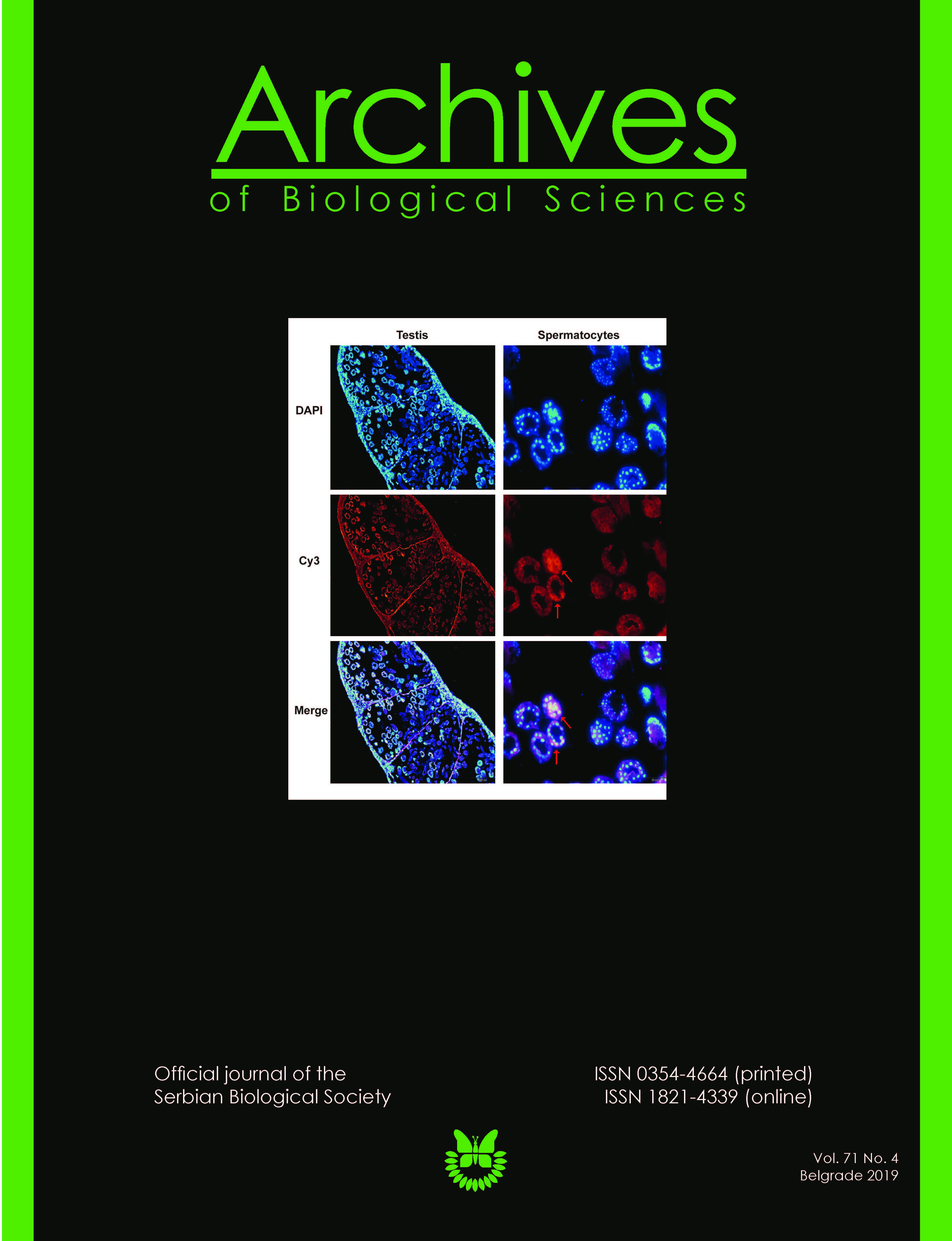
Disulfiram partially improves oxidative but not androgen status in rats exposed to cadmium
Keywords:
cadmium, disulfiram, oxidative stress, testes, ratAbstract
Paper description:
- The effect of disulfiram (DSF) on reproductive toxicity induced by cadmium (Cd) was studied.
- In the testes of Wistar rats, morphological, oxidative and testosterone changes were examined upon exposure to Cd and/or DSF.
- Cd triggered oxidative stress, affected the morphology of the testes and decreased testosterone production.
- DSF decreased the oxidative stress.
- DSF did not change the morphology of the testes and it did not affect plasma testosterone levels. Once impaired by Cd, Leydig cells could not be reactivated by DSF.
This article has been corrected. Link to the correction 10.2298/ABS200226010E
Abstract: We investigated the effect of disulfiram (DSF) on reproductive toxicity induced by subchronic exposure to cadmium (Cd). We examined the redox status and systemic testosterone changes in the testes and plasma of Cd-treated male Wistar rats. Rats were treated with 1 mg CdCl2/kg body weight (bw)/day (intraperitoneal administration) for 42 days; in the second experimental group, rats were given 178.5 mg DSF/kg bw/day by oral gavage for 21 days; in the third group, after administration of Cd for 21 days, DSF treatment was introduced on day 22 and lasted until day 42, with continuous Cd intake. Each experimental group had a matching control: untreated rats, rats that received for 21 days olive oil, the solvent for DSF; rats that started with olive oil intake from days 22-42. Exposure of rats to DSF modulated the oxidative status in the testes; thus, coexposure increased the Cd-induced reduction in total superoxide dismutase (tSOD), catalase (CAT), glutathione reductase (GR) and total glutathione-S-transferase (tGST) activities, and lowered the Cd-increased superoxide anion radical (O2●–) and malondialdehyde (MDA) concentrations. DSF did not affect testosterone production diminished by Cd, as Leydig cells, once impaired by Cd, could not be reactivated by DSF.
https://doi.org/10.2298/ABS190814057P
Received: August 14, 2019; Revised: September 4, 2019; Accepted: September 4, 2019; Published online: September 9, 2019
How to cite this article: Pavlović M, Đurić A, Stevanović I, Begić A, Vujanović D, Ninković M, Đukić M. Disulfiram partially improves oxidative but not androgen status in rats exposed to cadmium. Arch Biol Sci. 2019;71(4):747-53.
Downloads
References
Tchounwou PB, Yedjou CG, Patlolla AK, Sutton DJ. Heavy metal toxicity and the environment. In: Luch A, editor. Molecular, clinical and environmental toxicology. Vol. 3. Environmental Toxicology. Basel: Springer; 2012. p. 133-64.
Satarug S, Moore MR. Adverse health effects of chronic exposure to low-level cadmium in foodstuffs and cigarette smoke. Environ Health Perspect. 2004;112(10):1099-103.
Martelli A, Rousselet E, Dycke C, Bouron A, Moulis J-M. Cadmium toxicity in animal cells by interference with essential metals. Biochimie. 2006;88(11):1807-14.
Telisman S, Cvitković P, Jurasović J, Pizent A, Gavella M, Rocić B. Semen quality and reproductive endocrine function in relation to biomarkers of lead, cadmium, zinc, and copper in men. Environ Health Perspect s. 2000;108(1):45-53.
Pant N, Upadhyay G, Pandey S, Mathur N, Saxena D, Srivastava S. Lead and cadmium concentration in the seminal plasma of men in the general population: correlation with sperm quality. Reprod Toxicol. 2003;17(4):447-50.
Djuric A, Begic A, Gobeljic B, Stanojevic I, Ninkovic M, Vojvodic D, et al. Oxidative stress, bioelements and androgen status in testes of rats subacutely exposed to cadmium. Food Chem Toxicol. 2015;86:25-33.
Manna P, Sinha M, Sil PC. Cadmium induced testicular pathophysiology: prophylactic role of taurine. Reprod Toxicol. 2008;26(3-4):282-91.
Uygur R, Yagmurca M, Alkoc O, Genc A, Songur A, Ucok K, Ozen O. Effects of quercetin and fish n‐3 fatty acids on testicular injury induced by ethanol in rats. Andrologia. 2014;46(4):356-69.
Kitson T. Reinvestigation of the chemical reaction between disulfiram and ethanol. J Stud Alcohol. 1977;38(9):1771-2.
Johansson B. A review of the pharmacokinetics and pharmacodynamics of disulfiram and its metabolites. Acta Psychiatr Scand. 1992;86(S369):15-26.
Auclair C, Voisin E. Nitroblue tetrazolium reduction. In: Greenwald RS, editor. Handbook of methods for oxygen radical research. Boca Raton, Fl.: CRC Press; 1985. p. 123-32.
Sutherland MW, Learmonth BA. The tetrazolium dyes MTS and XTT provide new quantitative assays for superoxide and superoxide dismutase. Free Radic Res. 1997;27(3):283-9.
Li Y, Schellhorn HE. Rapid kinetic microassay for catalase activity. J Biomol Tech. 2007;18(4):185.
Habig WH, Pabst MJ, Jakoby WB. Glutathione S-transferases the first enzymatic step in mercapturic acid formation. J Biol Chem. 1974;249(22):7130-9.
Tietze F. Enzymic method for quantitative determination of nanogram amounts of total and oxidized glutathione: applications to mammalian blood and other tissues. Anal Biochem. 1969;27(3):502-22.
Ohkawa H, Ohishi N, Yagi K. Assay for lipid peroxides in animal tissues by thiobarbituric acid reaction. Anal Biochem. 1979;95(2):351-8.
Begic A, Djuric A, Gobeljic B, Stevanovic I, Lukic V, Stanojevic I, Ninkovic M, Saso L, Vojvodic D, Djukic M. The simple isocratic HPLC-UV method for the simultaneous determination of reduced and oxidized glutathione in animal tissue. Acta Chromatogr. 2017;29(1):67-84.
Jockenhovel F, Haase S, Hoermann R, Mann K. New automated direct chemiluminescent immunoassay for the determination of serum testosterone. J Clin Ligand Assay. 1996;19(2):138-44.
Lowry OH, Rosebrough NJ, Farr AL, Randall RJ. Protein measurement with the Folin phenol reagent. J Biol Chem. 1951;193:265-75.
Ruegg M, Meinen S. Histopathology in hematoxylin & eosin stained muscle sections Sop no MDC1A_M.1.2.004. Version 1.0 [internet]. Treat-NMD Neuromuscular Network; 2011 Apr 19 [updated 2014 Jan 28; cited 2019 Sep 09]. Available from: https://treat-nmd.org/wp-content/uploads/2016/08/cmd-MDC1A_M.1.2.004-68.pdf
Ognjanović BI, Marković SD, Ðorđević NZ, Trbojević IS, Štajn AŠ, Saičić ZS. Cadmium-induced lipid peroxidation and changes in antioxidant defense system in the rat testes: Protective role of coenzyme Q10 and Vitamin E. Reprod Toxicol. 2010;29(2):191-7.
Spiazzi CC, Manfredini V, da Silva FEB, Flores ÉM, Izaguirry AP, Vargas LM,Soares MB, Santos FW. γ-Oryzanol protects against acute cadmium-induced oxidative damage in mice testes. Food Chem Toxicol. 2013;55:526-32.
Saso L, Korkina L, Zarkovic N. Modulation of Oxidative Stress: Pharmaceutical and Pharmacological Aspects 2017. Oxid Med Cell Longev. 2017;2017.
Aitken RJ, Roman SD. Antioxidant systems and oxidative stress in the testes. Oxid Med Cell Longev. 2008;1(1):15-24.
Casalino E, Calzaretti G, Sblano C, Landriscina C. Cadmium-dependent enzyme activity alteration is not imputable to lipid peroxidation. Arch Biochem Biophys. 2000;383(2):288-95.
Dikalov S. Cross talk between mitochondria and NADPH oxidases. Free Radic Biol Med. 2011;51(7):1289-301.
Thévenod F. Cadmium and cellular signaling cascades: to be or not to be? Toxicol Appl Pharmacol. 2009;238(3):221-39.
Wang J, Zhang H, Zhang T, Zhang R, Liu R, Chen Y. Molecular mechanism on cadmium-induced activity changes of catalase and superoxide dismutase. Int J Biol Macromol. 2015;77:59-67.
Rahden-Staroń I, Grosicka-Maciąg E, Kurpios-Piec D, Czeczot H, Grzela T, Szumiło M. The effects of sodium diethyldithiocarbamate in fibroblasts V79 cells in relation to cytotoxicity, antioxidative enzymes, glutathione, and apoptosis. Arch Toxicol . 2012;86(12):1841-50.
Wadhwa S, Mumper RJ. D-penicillamine and other low molecular weight thiols: Review of anticancer effects and related mechanisms. Cancer Lett. 2013;337(1):8-21.
Djuric A, Begic A, Gobeljic B, Pantelic A, Zebic G, Stevanovic I, Djurdjevic D, Ninkovic M, Prokic V, Stanojevic I, Vojvodic D, Djukic M. Subacute alcohol and/or disulfiram intake affects bioelements and redox status in rat testes. Food Chem Toxicol. 2017;105:44-51.
Singh A, Kukreti R, Saso L, Kukreti S. Oxidative stress: A key modulator in neurodegenerative diseases. Molecules. 2019;24(8):1583.
Trachootham D, Lu W, Ogasawara MA, Valle NR-D, Huang P. Redox regulation of cell survival. Antioxid Redox Signal . 2008;10(8):1343-74.
Winterbourn CC, Hampton MB. Thiol chemistry and specificity in redox signaling. Free Radic Biol Med. 2008;45(5):549-61.
Zheng J, Lou JR, Zhang X-X, Benbrook DM, Hanigan MH, Lind SE, Ding WQ. N-Acetylcysteine interacts with copper to generate hydrogen peroxide and selectively induce cancer cell death. Cancer Lett. 2010;298(2):186-94.
Wu G, Fang Y-Z, Yang S, Lupton JR, Turner ND. Glutathione metabolism and its implications for health. J Nutr. 2004;134(3):489-92.
Jozefczak M, Remans T, Vangronsveld J, Cuypers A. Glutathione is a key player in metal-induced oxidative stress defenses. Int J Mol Sci. 2012;13(3):3145-75.
Ploemen J-PH, van Iersel ML, Wormhoudt LW, Commandeur JN, Vermeulen NP, van Bladeren PJ. In vitro inhibition of rat and human glutathione S-transferase isoenzymes by disulfiram and diethyldithiocarbamate. Biochem Pharmacol. 1996;52(2):197-204.
Townsend DM, Tew KD, Tapiero H. The importance of glutathione in human disease. B Biomed Pharmacother. 2003;57(3-4):145-55.
Giustarini D, Galvagni F, Tesei A, Farolfi A, Zanoni M, Pignatta S, Milzani A, Marone I, Dalle-Donne I, Nassini R, Rossi R. Glutathione, glutathione disulfide, and S-glutathionylated proteins in cell cultures. Free Radic Biol Med. 2015;89:972-81.
Downloads
Published
How to Cite
Issue
Section
License
Authors grant the journal right of first publication with the work simultaneously licensed under a Creative Commons Attribution 4.0 International License that allows others to share the work with an acknowledgment of the work’s authorship and initial publication in this journal.

